Production of Polyclonal and Monoclonal Antibodies
Introduction
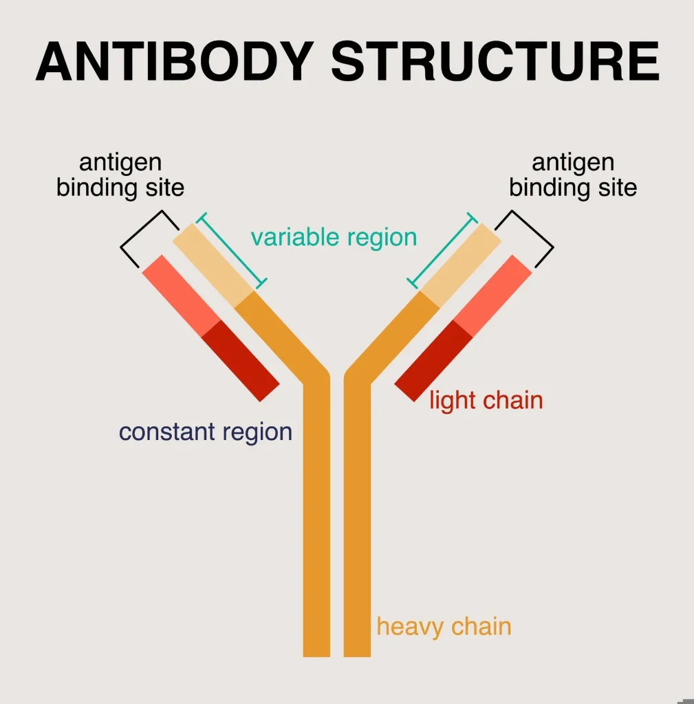
Antibodies are defensive proteins produced by the immune system in response to exposure to infectious microorganisms or vaccines. Their primary function is to recognize and bind specifically to antigens such as viruses, bacteria, or other foreign molecules. Once bound to the target, antibodies signal the immune system to eliminate the pathogen. In some cases, antibodies can directly neutralize microorganisms by blocking their replication or inducing their destruction.
Antibodies can also recognize foreign cells, such as those introduced during organ transplantation. In certain pathological conditions known as autoimmune diseases, antibodies mistakenly target the body’s own cells. Examples include systemic lupus erythematosus and multiple sclerosis.
Two major categories of antibodies are widely studied and used in biomedical research and medicine: polyclonal antibodies and monoclonal antibodies.
Polyclonal Antibodies
Polyclonal antibodies consist of a heterogeneous mixture of immunoglobulins produced by multiple clones of plasma cells. Plasma cells are differentiated B lymphocytes specialized in antibody secretion.
Because they originate from different B-cell clones, polyclonal antibodies recognize multiple epitopes on the same antigen. This multi-epitope recognition provides strong overall binding and increases sensitivity in many immunological assays.
Polyclonal antibodies are commonly used in:
- Immunological research
- Diagnostic assays
- Protein detection techniques
However, their heterogeneity can sometimes lead to variability between batches.
Monoclonal Antibodies
Monoclonal antibodies are identical immunoglobulins produced by a single clone of plasma cells. Unlike polyclonal antibodies, they recognize only one specific epitope on a given antigen.
Because of their high specificity, monoclonal antibodies are widely used in:
- Biomedical research
- Diagnostic testing
- Targeted therapies
- Oncology treatments
For example, if immune cells are exposed to a protein required for the proliferation of cancer cells, the produced monoclonal antibody can bind specifically to that protein. By blocking its function, the antibody can interfere with tumor growth without affecting other cellular proteins.
Key Immunological Concepts
Antigen
An antigen is any foreign substance capable of triggering an immune response.
Epitope
An epitope is the specific region of an antigen that is recognized by an antibody or by receptors on lymphocytes.
Oncology
Oncology is the medical discipline dedicated to the study, diagnosis, and treatment of cancer, including the molecular mechanisms underlying tumor development.
Hybridoma Technology
Hybridoma technology is the classical method used for the production of monoclonal antibodies. This technique allows the generation of immortal cell lines capable of producing a single type of antibody indefinitely.
The process relies on the fusion of antibody-producing B lymphocytes with immortal myeloma cells, resulting in hybrid cells known as hybridomas.
Hybridomas combine two important properties:
- Antibody secretion from B lymphocytes
- Unlimited proliferation from myeloma cells
General Workflow of Monoclonal Antibody Production
 The production process typically involves several major steps:
- In vivo immunization
- Preparation of myeloma cells
- Cell fusion
- Selection of hybridomas
- Screening of antibody-producing clones
- Cloning of selected hybridomas
- Large-scale antibody production and purification
1. In Vivo Immunization
Laboratory mice are the most commonly used animals for monoclonal antibody production. They are preferred because:
- Their genome shares approximately 95% homology with humans
- They are small and easy to maintain
- They reproduce rapidly and have a short lifespan
One frequently used strain is BALB/c, which is raised in controlled sterile conditions. Because these mice possess relatively low levels of natural antibodies, their immune system produces a strong response following antigen stimulation.
After immunization with the target antigen, the animal generates B cells capable of producing the desired antibody.
2. Fusion Partners
Two cell populations are required for hybridoma generation.
B Lymphocytes
B cells are isolated from secondary lymphoid organs such as:
- Spleen
- Lymph nodes
These cells include plasma cells capable of producing immunoglobulins and expressing the enzyme hypoxanthine-guanine phosphoribosyltransferase (HGPRT).
Myeloma Cells
Myeloma cells are immortal cancerous plasma cell lines that:
- Lack the HGPRT enzyme
- Do not produce immunoglobulins
Their main contribution to the hybridoma is unlimited proliferation capacity.
3. Cell Fusion Methods
After successful immunization, the animal is sacrificed and the spleen is removed under sterile conditions. Splenic cells (splenocytes) are then mixed with myeloma cells in a defined ratio.
Cell fusion can be achieved using two main techniques:
Chemical Fusion
Polyethylene glycol (PEG) is commonly used to induce the fusion of cytoplasmic membranes between the two cell types. PEG promotes high fusion efficiency. Dimethyl sulfoxide (DMSO) may be added to improve cell viability.
Physical Fusion
Electrofusion involves the application of electrical pulses that create temporary pores in the cell membrane, allowing cell fusion. This method is less destructive but more expensive.
Following fusion, excess fusion agent is removed by centrifugation and the cells are transferred into culture medium.
4. Selection of Hybridomas
The fused cells are cultured in a selective HAT medium containing:
- Hypoxanthine
- Aminopterin
- Thymidine
This medium allows only hybrid cells to survive.
Unfused myeloma cells cannot survive because they lack the HGPRT enzyme, while normal B cells have a limited lifespan. As a result, only hybridoma cells continue to grow.
Cells are distributed into multi-well plates to isolate individual clones.
5. Screening of Antibody-Producing Cells
Each positive culture well may contain hybridomas producing different antibodies. Therefore, specific antibody-secreting clones must be identified through screening assays.
Common detection methods include:
- Enzyme-Linked Immunosorbent Assay (ELISA)
- Radioimmunoassay (RIA)
- Hemagglutination tests
- Indirect immunofluorescence
These analyses are typically performed 9 to 11 days after cell fusion.
6. Hybridoma Cloning
Once antibody-producing hybridomas are identified, they must be cloned to ensure monoclonality. This step ensures that the antibody originates from a single cell clone.
Common cloning methods include:
Limiting dilution
Cells are diluted so that each culture well ideally contains a single cell. This technique is widely used because it is inexpensive, although it can be time-consuming.
Agarose cloning
Cells are cultured in a semi-solid agarose medium where they form visible colonies that can be isolated.
Flow cytometry sorting
This advanced technique allows rapid identification and isolation of hybridomas secreting the desired antibody from millions of cells.
7. Antibody Purification and Storage
Once produced, monoclonal antibodies must be purified before use. Several purification techniques are commonly applied:
- Gel filtration chromatography
- Ion-exchange chromatography
- Ammonium sulfate precipitation
The stability and storage duration of antibodies depend on their biochemical properties and storage conditions. Under appropriate conditions, antibodies can remain stable for several weeks to several years.